


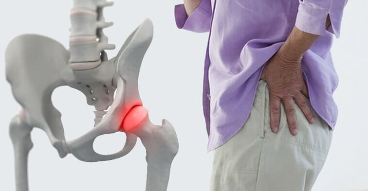
Pain in the hip jointthey are specific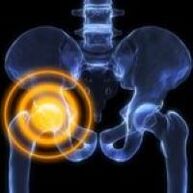 unpleasant and difficult to endure sensations caused by the pathology of the upper femur, acetabulum, nearby soft tissue structures. In terms of intensity, they range from weak to unbearable, in nature they can be dull, sharp, pressing, aching, explosions, perforations, etc. They often depend on load, time of day, and other factors. The causes of pain are determined using X-rays, CT, MRI, ultrasound, arthroscopy and other studies. Pain relievers and limb rest are recommended until diagnosis.
Causes of pain in the hip joint
Soft tissue injuries
The most common traumatic cause of pain is a contusion of the hip joint. Occurs when falling to the side or in direct impact, manifests itself in moderate acute pain, which quickly goes dull, gradually subsides and disappears within a few days, in severe cases - weeks. The support is preserved, the movements are slightly limited. Edema is detected locally, bruises are possible.
Injuries to the ligaments of the hip joint are rare, usually the result of traffic accidents and sports injuries, accompanied by severe pain, sometimes - a feeling of cracking (as from tear tissue). The pain decreases slightly, then often increases again due to edema. Swelling from the joint extends to the groin area, thigh.
The degree of dysfunction in the trauma of the ligamentous apparatus depends on the severity of the injury (strain, tear, rupture), which ranges from a slight limitation to the inability to support the leg. The pain increases with the deviation of the trunk, movements in the opposite direction to the damaged ligament.
Bone and joint injuries
Hip fractures usually occur in older people due to home or street trauma. A feature, especially in the presence of osteoporosis, is the absence of severe pain syndrome, mild edema. At rest, the pain is deep, dull, moderate or insignificant, with movements the painful sensations sharply increase. Support is sometimes maintained. A common symptom is the inability to lift a straightened leg from a prone position (a symptom of a stuck heel).
Transtrocanteric fractures are most commonly diagnosed in middle-aged and young people and develop as a result of high-energy trauma. Unlike cervical fractures, they are accompanied by an unbearable acute diffuse deep pain. Then the pain subsides, but remains very strong, difficult to bear. The joint is swollen, bruises are possible. Movement is severely limited. Support is impossible.
Isolated fractures of the greater trochanter are rare; they are found in children and young people; they are formed by a fall, a direct impact or a strong muscle contraction. The pain is acute, very intense, localized mainly on the external surface of the joint. Due to increased pain, the patient avoids active movements.
Hip dislocations occur during falls from a height, industrial and road traffic injuries, manifested in acute, unbearable pains that almost do not subside to reduction. The joint is deformed, the leg is shortened, bent at the knee joint, turned outward, less often inward (depending on the type of dislocation). Support and movement are impossible, when trying to move, the resistance of the spring is determined.
Acetabular fractures develop in isolation or are associated with hip dislocations. Characterized by acute explosive pain in the depths of the hip joint. Thereafter, the pain subsides slightly, but remains intense, preventing any movement. The leg is shortened, turned outward. Support is impossible.
Degenerative processes
With coxarthrosis in the initial stage, the pain is periodic, dull, of uncertain localization, appears at the end of the day or after a significant load, sometimes radiating to the hip, to the knee joint. At the beginning of the movements, a slight stiffness is possible which passes quickly. Subsequently, the intensity of pain increases, painful sensations are noted not only during movements, but also at rest. After intense exertion, the patient begins to limp. Movement is somewhat limited.
In severe coxarthrosis, the pain is deep, widespread, constant, aching, tortuous. Disturb both day and night. Stress resistance is reduced; when walking, patients lean on a cane. Movement is significantly limited, the affected leg is shortened, which leads to an increase in the load on the joint, increased pain when walking and standing.
Chondromatosis of the hip joint in its course resembles subacute arthritis. The pains are moderate, widespread, transient, combined with crunches, limitation of mobility. When intra-articular bodies are violated, blockages occur, characterized by intense sharp pain, impossibility or significant limitation of movements. After the cessation of the violation of the joint mouse, the listed symptoms disappear.
Trochanteritis usually forms with osteoarthritis of the hip joint, accompanied by an inflammatory-degenerative lesion of the tendons of the gluteal muscles at the point of their attachment to the greater trochanter, manifested by pain in the area of the lesion in the supine position on the affected side. There is increased pain when trying to abduct the hip with resistance.
Disorders of bone nutrition
Perthes' disease develops in children and adolescents, is characterized by partial necrosis of the femoral head, which is initially accompanied by a deep, dull, dull pain, sometimes radiating to the knee and hip. After a few months, the pain sharply intensifies, becomes constant, sharp, exhausting. The joint swells, movement is limited, and lameness occurs. Then the pain decreases, the degree of restoration of joint functions varies.
Downstream aseptic necrosis of the femoral head resembles Perthes disease, but is detected in adults, proceeds less favorably, in half of cases it is bilateral. At first, the pain is periodic, pulling. Then the pain syndrome intensifies, appears at night. At the peak of clinical manifestations, the pain is so intense that the person completely loses the ability to lean on the leg. Then the pains gradually subside. Movement limitations progress over approximately 2 years, resulting in osteoarthritis of the hip joint, contractures and shortening of the limb.
Solitary bone cysts form in the proximal metaphysis of the thigh in 10-15 year old boys, accompanied by intermittent non-intense pain in the hip joint. Edema is usually absent, with prolonged course contractures often develop, especially in young children. Due to mild symptoms, the cause of treatment is a pathological fracture or increasing limitation of movement.
Arthritis
Aseptic arthritis is manifested by undulatory pain in the joint, which increases in the early hours. The severity of pain ranges from insignificant to acute, strong, constant, significantly limiting physical activity. Stiffness, swelling, redness and local temperature rise are noted. Palpation is painful.
In rheumatoid arthritis, the hip joints are rarely involved, the lesion is symmetrical. Periodic pain first appears against the background of the change of seasons (autumn, spring), with a sharp change in weather conditions, during periods of hormonal changes after childbirth or during menopause. The pain is moderate or weak, diffuse, pulling or aching, markedly increased on palpation. It is combined with recurrent synovitis, edema, hyperemia, hyperthermia, increased limitation of mobility.
Infectious arthritis develops with the hematogenous or lymphogenic spread of infection, less often - with the penetration of the pathogen into the joint from neighboring tissues. Typically acute onset with rapidly increasing pain. The pain is intense, twitching, tearing, bursting, discomfort at rest, aggravated by movement, as a result of which the limb takes a forced position. Patients have fever, chills, sweating, severe weakness, edema, redness of the joint and increased local temperature.
In the absence of timely treatment, bacterial infectious arthritis can turn into panarthritis, a purulent inflammation of all tissues of the hip joint. It is characterized by a severe course with very acute diffuse shooting pains, frenetic fever, severe weakness, presyncope, significant hyperemia and hyperthermia.
Other inflammatory diseases
Upper thigh osteomyelitis can be hematogenous, post-traumatic, or postoperative. Hematogenic osteomyelitis is manifested by clearly localized, very sharp pain, spasms, tearing or dull, as a result of which the patient avoids the slightest movements of the limbs. There is a marked hyperthermia, severe intoxication.
Post-traumatic and postoperative osteomyelitis occurs with similar, but less pronounced symptoms. Typically, a more gradual onset against the background of an open fracture or postoperative wound, the appearance of purulent discharge. Pain in the hip joint increases within 1-2 weeks in parallel with the progression of signs of local inflammation.
Synovitis develops against the background of injuries, other diseases of the hip joint, less often it becomes a manifestation of allergies. In acute synovitis, the pain is usually mild, dull, explosive, gradually increasing due to an increase in the amount of intra-articular fluid. The joint is swollen, palpation is slightly painful, a symptom of fluctuation is determined. Chronic synovitis is asymptomatic, accompanied by a faint aching pain.
With intermittent hydroarthrosis, pain is also insignificant, accompanied by discomfort, limited mobility and disappears within 3-5 days after reverse resorption of the effusion. They are renewed at regular intervals, individual for each patient, and are caused by repeated accumulations of fluid in the joint.
Specific infections
Tuberculosis of the hip joint is a common form of osteoarticular tuberculosis, which is manifested by general weakness, fatigue, subfebrile condition. Then there are faint stretches or aching pains in the muscles, transient painful sensations in the joint when walking. The patient begins to spare the limb. As the pain progresses, they become moderate, diffuse, radiate to the knee, complemented by swelling, redness, synovitis. A protective contracture develops.
Joint pain, including the hip, can appear with brucellosis. In acute and subacute form, painful sensations that pull, twist, combined with periodic fever, lymphadenopathy, skin rashes. In a chronic course, the pain syndrome resembles that of aseptic arthritis, deformities are formed over time.
Congenital anomalies
Manifestations of hip dysplasia are determined by the degree of incongruity of the femoral head and acetabulum. With a complete congenital dislocation, pain appears immediately after the baby begins to walk, accompanied by lameness. With moderate subluxation, painful sensations occur at the age of 5-6, directly related to the load on the leg.
With a mild subluxation, the pathology is asymptomatic for a long time, the pain syndrome is manifested by the development of dysplastic coxarthrosis at the age of 25-30. The hallmarks of this arthrosis are the rapid intensification of pain, the early onset of pain at rest and at night, and the progressive limitation of movement. All forms of dysplasia are accompanied by asymmetry of the skin folds, the "click" symptom and limited mobility. In case of dislocation, shortening of the limbs is noted.
neoplasms
For benign neoplasms, a typical asymptomatic course. The pain is mild, intermittent, and often does not progress over the years. The growth of the tumor is accompanied by a slow increase in pain syndrome, recurrent synovitis. In the area of the hip joint, osteomas, osteoid osteomas, osteoblastomas, chondromas are most often detected.
Malignant neoplasms (osteosarcomas, chondrosarcomas) are characterized by the rapid progression of pain syndrome and other pathological manifestations. At first, the pain is minor, in the short term, without a specific localization, sometimes it gets worse at night. Subsequently, they become sharp, permanent, cutting, surrounding, spreading to the entire joint. The affected area is swollen, deformed. Weight loss, weakness, subfebrile condition are noted. With advanced neoplasms, painful and unbearable pains are eliminated only with narcotics.
Other reasons
Pain in the hip joint sometimes appears with lumbosacral plexitis and neuropathy of the sciatic nerve, however, they usually occupy an insignificant position in the clinical picture of the disease, fade into the background to intense pain on the back of the buttock and thigh, limb weakness and sensitivity disorders.
Pain syndrome of this localization is often detected in osteochondrosis and herniated disc. Pain can be detected with spondylitis, deformation of spondyloarthrosis and curvature of the spine. The pains are dull, periodic, aching, often intensifying during the period of exacerbation of the underlying disease. The cause of their appearance can be constant overload of the joint or the development of coxarthrosis.
Sometimes joint pain is triggered by a mental illness or a depressive disorder. Diabetes mellitus is often accompanied by enthesopathies, capsulitis, and other periarticular soft tissue injuries. Possible drug arthropathy when taking certain medications.
Diagnostics
In case of injuries, diagnostic measures are carried out by traumatologists. Degenerative and inflammatory diseases are managed by orthopedists and rheumatologists. In case of purulent processes, the participation of surgeons is required. The examination includes the collection of complaints, the study of the anamnesis, the physical examination, further research. Taking into account the peculiarities of the pathological process, the following methods can be used:
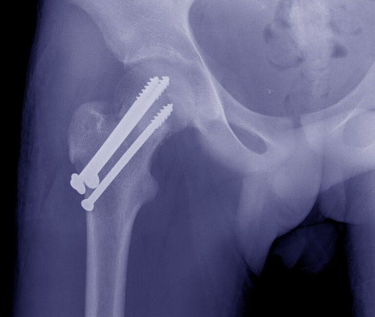
- X-ray. It is the basic technique for most joint diseases. It detects fractures, dislocations, changes in the contours of the acetabulum and the femoral head, marginal and intraosseous defects, bone growths, narrowing of the joint space.
- Ultrasound.More informative when studying soft tissues. It reveals signs of inflammatory and degenerative processes, areas of calcification. Used to diagnose effusion, joint mice.
- MRI and CT.Clarification techniques are used in case of ambiguous data from basic studies, to clarify the nature, prevalence and location of the pathological focus. Achievable with contrast.
- Puncture of the joint.It has a diagnostic or therapeutic and diagnostic character. It allows you to remove the effusion, study the composition of intra-articular fluid, determine the causative agent of infection by laboratory tests.
- Arthroscopy.During a visual examination of the joint, the doctor assesses the condition of the bone structures and soft tissues, if necessary, takes a biopsy sample for subsequent histological examination and performs therapeutic measures.
- Lab test.They are prescribed to determine signs of inflammation and markers of rheumatological diseases, to assess the general condition of the body, the activity of various organs in severe infectious or systemic pathologies.
Treatment
Help before diagnosis
In severe injuries, it is necessary to fix the joint by applying a splint from the foot to the armpit. In case of minor traumatic injuries, it is enough to provide rest to the leg. Cold is applied to the affected area. For severe pain, an analgesic is given. You can not make active movements with the limb, load the leg. It is strictly forbidden to try to eliminate the dislocation or displacement of the bones.
Tactics for non-traumatic diseases are determined by symptoms. With minor manifestations, it is allowed to ensure rest of the limb, the use of local remedies with analgesic and anti-inflammatory effects. In case of fever, weakness, severe pain, rapid growth of edema and hyperemia, it is recommended to immediately seek specialized help.
Conservative therapy
Dislocations are an indication for immediate reduction. In case of fractures, skeletal traction is usually applied, then patients are operated on or the limb is fixed with a plaster cast after callus marks appear. In elderly patients with hip fractures, immobilization with a derotation boot is allowed, which prevents rotational movements in the joint.
The rest of the patients are advised to relieve the hip joint. According to the indications, it is recommended to use orthoses or additional devices (crutches, cane). Prescribe massages, physiotherapy exercises, physiotherapy procedures:
- laser therapy;
- magnetotherapy;
- ultra-high frequency;
- ultrasound;
- electrophoresis with drugs;
- UHT.
It is possible to use NSAIDs, chondroprotectors, antibacterial drugs. Local agents are widely used. According to the indications, joint punctures, intra and periarticular blocks with hormones, intra-articular injections of chondroprotectors, substitutes of synovial fluid are performed.
Surgery
Interventions on the hip joint are performed with free access and with the aid of arthroscopic equipment. Taking into account the type of pathology, the following can be performed:
- Traumatic injuries:open reduction of hip dislocation, acetabulum reconstruction, neck osteosynthesis, trochanteric fractures.
- Degenerative processes:arthrotomy, arthroscopy, removal of free intra-articular bodies.
- Tumors:removal of neoplasms, bone resection, disarticulation of the hip joint, abdominal amputation, abdominal abdominal resection
In case of contractures, ankylosis, scarring of the periarticular tissues, repair, arthroplasty and arthrodesis are performed. Endoprosthesis is an effective way to restore limb functions in diseases of various origins, accompanied by limitation of movement or destruction of the joint.